What is Myasthenia Gravis?

Myasthenia gravis (MG) is a neuromuscular condition in people and animals that causes a weakness of the skeletal muscles.  Often, this weakness is exercise-induced and with short periods of rest the animals can recover a modicum of strength and movement.  The disease is considered a 'junctionopathy' because the synapse, or junction, of the nerve and muscle is the part of the pathway that is affected.  In particular, it is the receptor on the muscle side of the junction that is unable to function properly.  Basically, the muscle can't 'hear' the nerve talking to it.

MG can be an acquired disease or present at birth, congenital.  Congenital MG is uncommon in veterinary medicine.  We typically see the acquired form, so that's what we'll focus on in these blogs.

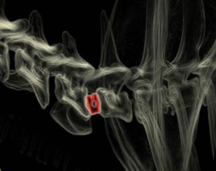
A radiograph (X-ray) of a dog with generalized megaesophagus.  The blue arrows show the outline of the dilated esophagus.

Acquired MG is recognized in 3 forms in veterinary medicine.  There is a generalized form that leads to the exercise induced weakness, a focal form that often affects the muscles of the esophagus, and a fulminant form that causes profound weakness of the respiratory muscles and can lead to death without life support and immediate therapy.  While patients often respond well to medical therapy and their strength usually improves dramatically, many veterinarians consider involvement of the esophagus to be a bad sign, even when it's only the focal form of the disorder.  That's because changes in the esophagus lead to regurgitation and might cause aspiration pneumonia.  Aspiration pneumonia is difficult and often very expensive to treat!

This cartoon depicts the antibody blocking the receptor that leads to the muscle weakness

In terms of how the disease comes to be, it is an autoimmune disorder.  For some reason we do not understand, the patient's own body begins making antibodies against the muscle receptor.  This receptor is called the Nicotinic Acetylcholine Receptor.  The antibodies then prevent the chemical signal from the nerve (acetylcholine) from being able to bind to the receptor and transmit the signal from the nerve to cause the muscle to contract.  At the level of the junction, or synapse, it becomes a competition between the antibody and the acetylcholine to inactivate or activate the receptor respectively.

In the next blog post, we'll discuss some of the ways we diagnose this disease, and we'll follow that one up with a discussion on therapy and resolution...

What is Veterinary Medical Acupuncture?

Acupuncture has been used for centuries in Asia to treat a variety of ailments.  In some versions of this ancient practice, small needles are placed in the skin with the goal of manipulating Qi (or Chi).  Recently, researchers have begun to investigate these techniques using scientific methods and tools more commonly associated with what is considered 'Western Medicine'.  What we're learning is fascinating!  And it has lead to the development of techniques that are referred to as Medical Acupuncture.

Over thousands of years of observation, what began as a technique for bloodletting was refined into the modern version of acupuncture.  When one looks closely at the places that are used for 'needling', you can see why these points were chosen.  There are often blood vessels and nerves traveling through the area just beneath the skin!

Medical Acupuncture attempts to combine evidence-based 'western' medicine or scientific techniques with the art of acupuncture.  This school of thought doesn't subscribe to the concept of Qi for developing a treatment plan.  Rather, it uses the neuroanatomical structures and relationships that are present at the sites to functionally interact with the nervous system to affect other organs and body systems.

We now know that the appropriate use of acupuncture needles can make major changes in the spinal cord's intrinsic 'wiring' or 'programming'.  We can actually use these needles to 'wind down' the spinal cord in situations related to the perception of chronic pain.  What's more, by reinforcing the signalling pathways in the spinal cord with acupuncture, there are patients that have suffered spinal cord injuries that can gain significant benefits.  And, in the case of peripheral nerve diseases, we're seeing similar results!  In the end, medical acupuncture is based on ancient techniques that are very safe, use no drugs, and have virtually no side-effects.  

Dr. King recently completed a rigorous certification process in Medical Acupuncture for Veterinarians, and we've added it to our armament of tools here at LOVN to help those patients that might have limited or no other options.  If you have questions about acupuncture or how we practice acupuncture for your pets, please feel free to contact us.  We're very excited to be able to offer these techniques to your pets and eager to discuss what we're learning and how it's helping those we might not have been able to help otherwise!

What is Canine Cognitive Dysfunction?

Did you know that dogs can develop a condition that is startlingly similar to Alzheimer's Disease in people?  It's true.  It's called Canine Cognitive Dysfunction (CCD) and a large percentage of our best friends will begin to show signs of this condition as early as 6 years old!  

Diagnosis of CCD is difficult.  There are sometimes changes on an MRI, but at other times all the diagnostic tests are normal.  That's why we, as veterinarians, rely so much on you, the pet owner, to identify signs of this condition and begin to intervene as soon as we can.  

You see, just like with Alzheimer's Disease, there is no cure for CCD.  There are lots of supplements, vitamins, botanical products, and even medications that can be used to slow the progression of the disease, but no one knows how to reverse the effects.

This particular disease is one that has caused me very personal heartbreak.  I had to say goodbye to the best-dang-dog-I've-ever-known, Scout, because of it.  

Professor Stout Scout in his prime ...

Professor Stout Scout in his prime ...

I met Professor Stout Scout when I was a technician working at an emergency veterinary clinic in Charleston, SC.  He came in the clinic as a stray and left that same night as my best friend and constant companion.  He went with me to Athens, GA for vet school; to Blacksburg, VA for my internship; and all the way to Davis, CA for my residency.

It was in California that I first started to notice some of the telltale signs of CCD.  Veterinarians use the acronym D-I-S-H to describe these signs.  

Disorientation is common.  

Interactions with their owners or other pets in the household may change

Sleep/Wake cycles are often disturbed

House-training can be lost as well

Scout started to show all these signs, and it seemed to be progressing rather rapidly.  Well, I'm insatiably curious and I had to know if my buddy was starting to show signs of CCD, or if there was something else going on.  Because brain tumors and other diseases can cause these signs too.  So Scout had an MRI and a spinal tap at UC Davis.

Typical structural changes in a dog's brain with CCD on MRI include loss of brain tissue, dilation of the fluid-filled ventricles, and shrinkage of the interthalamic adhesion.

Typical structural changes in a dog's brain with CCD on MRI include loss of brain tissue, dilation of the fluid-filled ventricles, and shrinkage of the interthalamic adhesion.

He must have read the same text books I was studying at the time, because he even had the structural brain changes that happen with CCD.  So, he'd checked all the tick-boxes for me.  I went on a search for something that might help.

I tried everything, and in the end found a cocktail of supplements and vitamins that seemed to slow his decline.  I was able to get another couple of years or so with him, but CCD won in the end.  Scout's quality of life declined, and I finally had to make that difficult, heartrending, and compassionate decision to help him slip off this mortal coil with all of the dignity and peace that I could muster for him.

So, if you think your older pet is starting to show signs of CCD, contact your veterinarian to have a discussion about the signs you're seeing.  We still can't reverse the signs of this disease so early intervention is key...

The Trifexis Debate

Working as Neurology technicians, we are regularly asked questions from owners pertaining to the daily care and maintenance of their beloved pets. These questions range from diet standards to bowel movement regularity. One question I have commonly been asked since working at Live Oak Veterinary Neurology is regarding the safety of using Trifexis. I believe that the majority of canine owners are aware of, or heard about, the WSB-TV Atlanta story by Jim Strickland linking several canine deaths to the use of Trifexis. My intent of this blog is to discuss our practice’s views on the use of Trifexis.

In order to write a well-informed blog on this topic, I reached out to several representatives of Elanco, producers of Trifexis. Through multiple conversations, assessment of FDA research, and discussing the topic with Dr. King, it is our practice’s opinion that Trifexis is a safe and effective tool in the fight against those pesky fleas and deadly heartworms.

Visit the American Heartworm Society website to learn more about these deadly parasites.

Visit the American Heartworm Society website to learn more about these deadly parasites.

With that said, when a client consults with our neurologist about their pet’s epilepsy, we recommend they not use Trifexis or Comfortis. The reason being, when we manage seizures, we want to be able minimize any external variables that could affect the pet’s symptoms. The component of Trifexis that controls fleas, spinosad, also the main drug in Comfortis, crosses the Blood Brain Barrier (BBB). The BBB’s sole purpose is to control what chemicals can get into the brain. With spinosad’s ability to cross the BBB, its presence is one factor in the patient’s epileptic mystery that we can control. Seizures are a brain disease so controlling what comes and goes is our main objective.

According to the research conducted to get the medication approved by the FDA, giving Trifexis or Comfortis does have its risks, just like any other medication out there. The most commonly seen side effect is vomiting. Many times this can be alleviated by just adjusting the way that you give the medication. I strongly recommend that, if you are seeing these side effects, contact your veterinary team or Elanco directly on their Technical Support line.

For questions regarding the use of Trifexis or Comfortis, please contact Elanco at 1(888)545-5973 or visit their website at http://www.trifexis.com/safetyfacts/.

If you feel that your pet is experiencing a side effect of Trifexis or Comfortis, please contact Technical Support at 1(888)545-5973 and speak with the company’s technicians or veterinarians. 

What is the Treatment for Chiari-like Malformation (COMS)?

I often describe the disease process involved in these cases using the analogy of putting your finger over the end of a garden hose.  When you do, the same volume of water comes through as it did without your finger in place, but the aperture is much smaller.  This causes changes in the hydrostatic pressure of the flow through the end of the garden hose.  

With every heartbeat, there is a sudden surge of fluid through the hole at the base of the skull.  This allows the skull to accommodate the increased volume of blood that comes in with every heartbeat. But the change in pressure that happens with the cerebellum herniated (or just crowded) at the base of the skull is just like having your thumb over the end of the garden hose ...

This is a video of a special MRI sequence performed in a human that shows the pulsatile flow of spinal fluid caused by the heartbeat ...

Management of this condition with medications (like prednisone and omeprazole) attempts to reduce the production of spinal fluid.  This is analogous to turning down the spigot while keeping your thumb over the end of the garden hose ...


Surgery removes the compression on the cerebellum to allow the fluid to pass through more easily.  This is akin to taking your thumb off the garden hose ...


I mention all of this, because I want to remind everyone that surgery doesn't directly make the syringohydromyelia (SM) in the spinal cord go away.  Rather, it prevents the disease from getting worse by removing the underlying cause.  That's important because it plays into how we define 'success' when looking at surgery.

I always caution my clients to think of a successful surgery as - a patient that recovers from the procedure with no complications and does not get worse over time due to progression of the SM.  In other words, these patients may still require medications and the surgery does not (necessarily) make the SM go away.  If we get improvement beyond this definition of success, then I call that 'icing on the cake'.  Using this definition of success, we are successful with surgery more than 80% of the time.  

Most of the surgical failures happen months to years after the procedure due to scar formation at the surgical site.  This seems to be where the titanium mesh has its greatest benefit.  Since I've been using the mesh, I haven't had any patients worsen in that time frame.  

Also, when we remove the bone, there is still a thick membrane (the meninges and particularly the dura mater) that restricts and confines the cerebellum and brain.  By patching in a piece of tissue, we can make more room within this membrane and relieve the compression.  It's very similar to letting out the waist in a pair of pants.  To help prevent scar tissue from forming here and undoing all that we've done, I use tissue collected from the patient's themselves (autograft) rather than material that might cause more inflammation or rejection of the graft.

What is Cervical Disc Arthroplasty (CDA)?

Cervical Disc Arthroplasty (CDA) is a relatively new procedure that is used to treat Disc Associated Wobbler's Syndrome (DAWS).  It was developed by Dr. Filippo Adamo ECVN, the inventor of the artificial disc implant that we use in the procedure (The Adamo Spinal Disc).  Dr. Adamo created the technique based on disc replacement surgeries that are done in people because of the tendency for DAWS patients to develop a 'domino lesion' of DAWS in the adjacent discs shortly after surgical correction of the first lesion.

This is a picture of the instrument table and set-up before placing the disc implant in a patient.

This is a picture of the instrument table and set-up before placing the disc implant in a patient.

The disc implant serves two purposes in treating DAWS.  First, it distracts the intervertebral space to relieve the compression on the spinal cord.  This is similar to the stated goal-of-therapy of several of the older procedures as well.  But where the CDA procedure differs, is in it's ability to maintain movement and range-of-motion at the surgical site.  By maintaining the dynamic nature of the intervertebral disc space, the CDA technique seeks to minimize the effects on the adjacent discs and prevent the dreaded 'domino lesion'.

We can place more than one disc implant in a patient when indicated.

We can place more than one disc implant in a patient when indicated.

The Adamo Spinal Disc is currently in it's 5th iteration.  The current design uses state-of-the-art manufacturing techniques and materials to improve the device and the surgical outcome for our patients.  


This is the complete Adamo Spinal Disc implant.  The white material on the back of the implants promotes bony fusion between the vertebrae and the implant.

This is the complete Adamo Spinal Disc implant.  The white material on the back of the implants promotes bony fusion between the vertebrae and the implant.

First, the device is made of surgical grade titanium.  This lightweight and durable metal is also MRI safe.  Which means that we can safely repeat MRI studies of the patients if needed after the surgery.  In fact, repeat MRI studies are part of our post-operative evaluation for this procedure.  Because the technique is so new and cutting edge,  LOVN performs these post-operative MRI studies for these patients at no cost in exchange for using their outcomes and data for future studies and scientific papers.  That's how much we believe in this procedure!

Second, the device uses a small thermoplastic articular surface to maintain the movement at the site.  It works just like a 'ball and socket' to allow the two parts of the implant to move relative to one another.  If the contact surfaces were made of metal, small microparticles would be produced from the friction and could lead to worsened inflammation or other problems.

Here you can see the two parts of the Adamo Spinal Disc and the 'ball and socket' articulation between the two.  You can also see the screw threads that allow us to remove the positioning pins once the implant is in place.

Here you can see the two parts of the Adamo Spinal Disc and the 'ball and socket' articulation between the two.  You can also see the screw threads that allow us to remove the positioning pins once the implant is in place.

Finally, the surface where the implant contacts the end plate bone of the vertebrae is specially treated in two ways to help in the healing process.  First, the surface is treated in a very special way to create titanium 'pores' or tunnels that encourage bone growth into the implant.  Once the bone grows into these 'tunnels' the interface between the bone and the implant becomes incredibly strong and durable.  Secondly, the titanium porosities are coated with hydroxyapatite crystals.  This is the same material that your body produces to create bone.  It serves as a substrate to speed up the bone growth into the pores and the ultimate incorporation into the vertebrae.


What is a CUSA?

CUSA is the acronym for cavitronic ultrasonic surgical aspirator.  This is an amazing device that is used in tumor surgery in a select number of hospitals around the world, and even fewer veterinary hospitals!

The CUSA uses ultrasonic frequencies to basically emulsify the target tissues.  The ultrasound waves cause the cells to dissociate from one another while the device irrigates the area with sterile saline.  The same handpiece then aspirates the fluid back out of the surgical site.  Because the device can be fine-tuned, it allows the surgeon to remove tumors with minimal effect on the adjacent tissues.  It can even 'skeletonize' the blood vessels which massively decreases the intraoperative bleeding!

We use our CUSA for removal of brain tumors, spinal cord tumors, and other delicate procedures involving the central nervous system.  Our SonoCure is the premier CUSA developed exclusively for veterinary use.  One of the most amazing parts of this device is the bone tip.  By using an exclusive technique of delivering longitudinal-torsional movement, the bone handle can emulsify (or liquify) bone in very delicate areas while protecting the vital tissues just next to it!

The bone tip uses a special longitudinal-torsional movement to add unparalleled precision to removal of bone immediately adjacent to delicate and vital tissues.

The bone tip uses a special longitudinal-torsional movement to add unparalleled precision to removal of bone immediately adjacent to delicate and vital tissues.

The addition of the SonoCure CUSA to our equipment at LOVN is just another way that we strive to provide the very best in medical and surgical outcomes for you and your pet!  To remain on the cutting-edge of surgical technique and technology is our promise to you.

What is Caudal Cervical Spondylomyelopathy (CCSM)?

This blog post is going to discuss the second form of "Wobbler's Disease" in dogs, Caudal Cervical Spondylomyelopathy or CCSM.

The breeds most commonly affected with CCSM include the Mastiffs and the Great Dane, but almost any large breed dog can be affected.  Typically, this condition is seen in younger dogs as it is a developmental problem (contrasted with the degenerative problem that causes Disc Associated Wobbler's Disease or DAWS).  

Again, to contrast with DAWS, the compression of the spinal cord is due to bony changes rather than discs, tendons, ligaments, or soft tissues.  The parts of the vertebrae that form the bony tunnel of the vertebral canal develop abnormally and are thickened towards the tail-end of the vertebrae.  This bony thickening is most noticable along the lateral (side) walls of the tunnel called the pedicles.  So, the compression is from the side in CCSM, whereas it's mostly top-to-bottom with DAWS.

Once again, there are two different options for therapy, medical management vs surgery.

With medical management, the goal is to control the dog's clinical signs.  But the drugs don't do anything to stop the disease progression.  And, sometimes, the disease can progress very suddenly due to vascular or 'bruising' injuries to the spinal cord!  Othertimes, the progression is slow and due to progressive bony changes at the true joints in the vertebrae, the facets.

Thickening of the facets can often cause progressive signs in CCSM due to worsening spinal cord compression...

Thickening of the facets can often cause progressive signs in CCSM due to worsening spinal cord compression...

Surgery attempts to alleviate the compression and works with medical management to control the clinical signs but also to halt the progression of the disease.  In the ideal situation, surgical decompression even allows us to stop the medications.  

Unlike DAWS which can suffer a 'domino effect' following surgery, this isn't common with CCSM decompression.  But, just like DAWS, there are several surgical options for pets with CCSM.  Some include decompression by removing bone from the top and sides of the vertebral canal.  Others involve a distraction-fusion technique to allow the bone to resorb on its own and alleviate the compression.

Diagnosis of this disease often requires an MRI or a CT scan combined with a myelogram.  In the end, the diagnosis will play an integral role in deciding which medical or surgical option is right for each individual patient.

What is Chiari Malformation (COMS)?

Chiari malformation is the term used in human medicine that equates in veterinary medicine to Caudal Occipital Malformation Syndrome, COMS.  The most commonly affected breed with COMS is the Cavalier King Charles Spaniel.  With the advent of MRI we are finding this condition in several other breeds as well such as the Chihuahua, Yorkshire Terrier, and other small breeds of dogs.  

COMS is a condition where the base of the skull in the dog is abnormally shaped.  This leads to a more 'rounded skull' and, in turn, causes the part of the brain at the base of the skull (the cerebellum) to be 'crowded'.  Because this skull defect is present at birth and through the growth phase of the puppy, once the dog reaches adulthood, the skull changes are static and don't change.  But the after-effects are progressive.

This is a sagittal MRI of a dog with COMS.  You can see the cerebellum at the base of the skull has slipped out of the base of the skull into the 1st vertebrae of the neck in what is termed a cerebellar herniation.  The SM secondary to the…

This is a sagittal MRI of a dog with COMS.  You can see the cerebellum at the base of the skull has slipped out of the base of the skull into the 1st vertebrae of the neck in what is termed a cerebellar herniation.  The SM secondary to the COMS is present as the white area in the spinal cord just behind the cerebellum.

You see, COMS is associated with another condition that takes place in the spinal cord 'downstream' from the skull in the dog's neck.  This condition is called syringohydromyelia or SM.  SM is a progressive degenerative condition in the spinal cord that can happen anytime the flow of spinal fluid is disrupted.  

Scoliosis, or a left-right bending malformation f the spinal cord can also happen in cases of COMS and SM

Scoliosis, or a left-right bending malformation f the spinal cord can also happen in cases of COMS and SM

Most people know that the spinal cord has a left and right side that correspond directly to the left and right sides of the body.  If you damage the left side of the spinal cord, the signs associated with that damage will be seen on the left side of the body.  However, did you know that the spinal cord also has a 'front' and 'back' that carry specific types of information?  

The part of the spinal cord that is towards the chest of a person or animal carries information from the brain to the body, instructions on how to move.  The part of the spinal cord towards the back of the person or animal carries information from the body to the brain, sensory information if you will.

Interestingly enough, SM almost always forms in the sensory part of the spinal cord.  So it leads to abnormal sensations rather than weakness or abnormal movments.  That means that dogs with SM manifest some very odd and quite varied behaviors secondary to the abnormal sensations that their spinal cord is causing them to feel.  Imagine, that every time you got excited your toes began to itch and burn, your ear began to feel hot, or your arm became numb and tingly...

The most striking manifestation of this abnormal sensation is called 'phantom scratching'.  In this situation, dogs will begin to scratch 'at' their ear, but just a little bit off to the side.  Almost as if they were scratching the air!

Diagnosis of COMS and SM requires an MRI.  No other tests are able to definitively diagnose this condition.  There are other tests that have been used to support the diagnosis though, including BAER hearing tests, CT scans, ultrasound, and even infrared imaging.  

Treatment for COMS is sometimes medical, and sometimes surgical.  There are several drugs that can be used to help control the condition, and surgery can be very successful (depending on how one defines 'success').

What is Disc-Associated Wobbler's Disease (DAWS)?

I mentioned in an earlier post that the term "Wobbler's Disease" is actually used to describe two different conditions in dogs, Caudal Cervical Spondylomyelopathy and Disc Associated Wobbler's Disease.  So I figured we would use these next few blog posts to cover these conditions in a little more detail.  Today we'll discuss Disc Associated Wobbler's Disease or DAWS...

DAWS is a condition that we see most often in older large breed dogs.  The Doberman Pinscher is the most commonly affected breed.  With this condition, slow degeneration of the intervertebral discs in the neck lead to thickening of other ligaments around the spinal cord and bulging of the disc itself.  This causes a progressive compression of the spinal cord, typically in the middle-to-lower region of the neck.

                         Guilty as charged ... Dobermans are the most common breed to be affected by DAWS....

                         Guilty as charged ... Dobermans are the most common breed to be affected by DAWS....


Compression of this part of the neck can lead to deficits in the forelimb reflexes and weakness.  This part of the disease manifests as short and choppy steps with the forelimbs.  But the nerve fibers carrying information to the rear limbs are also affected often without any changes in the reflexes.  The rear limbs, then, develop a long a loping gait that oversteps the normal stride and almost looks like the limbs are 'floating'.  Combine the two very different strides, and we see what is sometimes called a 'two-engine gait'.

This video is an example of the 'two-engine gait'.  It's been supplied courtesy of Dr. Michael Wong DACVIM (Neurology)  at Southeast Veterinary Neurology in Miami, FL.


There are two major schools of thought when it comes to treating these patients.  The first group recommends medical management and surgery as a last option.  The second group is more pro-active with surgical intervention.  At LOVN we use both medical and surgical management depending on the severity of the disease and other factors.  Every case is unique and deserves a unique approach...

Why would one recommend medical management over surgery for a condition that is obviously due to a structural problem that surgery might correct?  Excellent question!

You see, there was often a complication with surgery called a 'Domino Effect'.  Many surgical techniques used different approaches to either relieve the compression by removing the disc and/or other compressive soft tissues.  Sometimes, the disc was removed, and the disc space was widened by placing a 'spacer' between the vertebrae.  This would then lead to fusion of the two vertebrae (a distraction-fusion technique).  In some cases this was a bone graft taken from another dog, and in other cases it was a metal implant.

Some people propose that the affected disc space would then lose it's range-of-motion from either a successful surgical fusion or from the degeneration and collapse of the disc itself.  Biomechanically, this would cause the adjacent discs to have to take on greater degrees of movement for the animal to maintain its overall range of motion in its neck.  Have you ever taken a moment to think about all the different ways you can move your neck?  It's truly remarkable!

In theory then, the additional work required of the adjacent discs would cause those discs to degenerate as well and the whole process would start all over, just next door....

Still, the concept of 'distraction' is often considered important, because the compression of the spinal cord isn't just due to the bulging disc.  There are ligaments on top of the spinal cord that become thickened in response to what the body perceives as an instability.  Removing or addressing the bulging disc doesn't directly impact these tissues.  But with distraction, the ligaments are pulled taught and the compression is lessened.  Maintaining this distraction, then, might allow those thickened ligaments to thin out again and remove the compression from that side without a second procedure.  

Another new technique called the Cervical Disc Arthroplasty uses a two-part implant to replace the intervertebral disc.  This device is designed to maintain the distraction at the disc space but still allow movement.  The thought here is that maintaining movement at this disc space could possibly reduce or eliminate the dreaded Domino effect.

The Adamo Spinal Disc replaces the affected disc, but because it is composed of two moving parts, it maintains both movement and distraction at the surgical site.

The Adamo Spinal Disc replaces the affected disc, but because it is composed of two moving parts, it maintains both movement and distraction at the surgical site.

Finally, some surgeons attempt to remove the compression from the 'top' or the 'back of the neck' with a procedure called a Continuous Dorsal Laminectomy.  The theory behind this technique is that it removes the compression from the top of the spinal cord and allows the spinal cord to 'float' over the bulging disc.  Also, because it doesn't involve the affected disc, some motion can be maintained at this site, which also might reduce the chances of the Domino effect.

With so many options, and the cause of the 'domino effect' unclear, it's impossible to make a standard recommendation for all dogs with DAWS.  That's why it's important to have a team of professionals like your regular veterinarian and LOVN to help guide you in your choices.

What is Feline Infectious Peritonitis (FIP)?

Feline Infectious Peritonitis (FIP) is an infection caused by a virus in cats.  It is a very unique and complicated infection that is dependent on several factors involving the viral strain and the host itself.  Many years of study and research went into uncovering this strange viral infection and understanding how it happens, but much is still unknown and there is no known effective therapy.

FIP starts as a relatively simple Feline Coronavirus.  This is a type of virus/infection that can happen in many different animals with viruses that are often specific to that particular species.  Typically, the coronavirus causes diarrhea and gastrointestinal signs and is self-limiting.  Meaning, most animals recover from the infection without intervention or consequence.

But in some cats, some viral strains are able to transform into FIP.  The virus is still a coronavirus so testing for these viruses will often still give a positive result.  But the FIP strains take on new properties and abilities that allow them to infect the white blood cells that would normally kill them and clear the infection.  This allows the FIP strain to survive past the host's initial disease state and GI signs and evade destruction by the immune system.

The next step in the evolution of FIP as a disease can take place months to years after the initial infection and can come on with no warning.

FIP hiding in the immune system itself causes inflammation that begins to damage the host's own body.  There are two ways that this can happen.  Depending on what type of inflammatory response takes place, a cat can develop FIP as a 'wet' or a 'dry' form.  In cases of neurologic disease, the 'dry' form is the more common of the two.

In 'wet' form FIP, antibodies and inflammation due to the virus begins to irritate the blood vessels of the body.  This irritation leads to the vessels becoming 'leaky' and fluid will begin to infiltrate into the body cavities.  This fluid has very typical characteristics and can cause abdominal swelling and difficulty breathing due to the abdominal cavity or the chest becoming filled with fluid.

In the 'dry' form of FIP, the infected white blood cells begin to coalesce into clumps of cells called granulomas.  These granulomas can settle out into any of the body's tissues and begin to cause organ damage.  In the brain, the most common place to find them is the coverings of the brain (meninges) and the lining of the fluid filled spaces inside the brain itself (the ventricular system).  In fact, there is a very common MRI finding in cats with FIP that shows marked inflammation of a particular part of the ventricular system called the mesencephalic aquaduct or Aquaduct of Sylvius.

An MRI image of a cat as if the cat were looking out of the screen at the viewer.  The bright lines outline the ventricles (normal fluid filled spaces in the brain).  But the ventricles are swollen and dilated from the disease.  The l…

An MRI image of a cat as if the cat were looking out of the screen at the viewer.  The bright lines outline the ventricles (normal fluid filled spaces in the brain).  But the ventricles are swollen and dilated from the disease.  The lining is bright white because of the inflammation or granuloma formation secondary to the virus.

The prognosis for cats with FIP regardless of the form it takes, wet or dry, is guarded to grave.  There are medications that can be helpful for short periods of time, but there is no way to clear the virus from the affected pet.  Diagnosis is sometimes easy and at other times requires advanced testing and ruling out a multitude of other diseases.

What are seizures in dogs and cats and how are they treated?

A seizure is a symptom of certain diseases affecting the brain.  In medical terms, it's also referred to as 'hypersynchronous cerebrocortical discharge'.  That's because, in a healthy animal, the cells in the brain send electrical signals over relatively long distances and don't 'fire' all at once in any particular area.  On an EEG, this looks like little squiggly lines in health, but if enough cells fire together (hypersynchronous discharge) it looks like a big 'spike' on the EEG readout.

This is our Electroencephalogram in action ...

This is our Electroencephalogram in action ...

Like we said, seizures are a symptom of disease.  The most common disease to cause seizures in dogs is Idiopathic Epilepsy.  But liver disease, kidney disease, changes in calcium or sodium in the blood, even brain tumors and auto-immune diseases can cause seizures.  That's why we often recommend an MRI and a spinal tap to look at your pet's brain and determine the underlying cause.

Still, seizures as a symptom (even if the underlying cause isn't progressive - like Idiopathic Epilepsy or a 'stroke') are often progressive.  That means that, the more a pet has seizures, the more likely they are to have more seizures and the more severe the seizures are likely to become.

For this reason, we treat seizures to maintain a certain level of control.  While it is possible to make seizures go away completely; in many cases, the adverse effects of the drugs make this a terrible option.  You see,  the more we give drugs to control seizures, the more we sedate the patient.  If we go to far with the medications, there won't be any seizures but your pet might be mistaken for a doormat!

There are many different options for treating seizures in our pets.  First, we attempt to identify and treat the underlying cause of the seizures.  Then we try to find the right drug or combination of drugs for that particular pet and your particular situation.  That's why we call it the 'art of seizure management'.

What does the 'strength' of the MRI magnet mean?

MRI magnet strength is measured in a scientific unit called a Tesla.  For short-hand we use the abbreviation 'T' for this.  So a 1.5T magnet is one-and-a-half-times stronger than a 1.0T magnet.  There are MRI's used in experimental research that are 17.0T as of 2014 and there are even stronger magnetic fields that have been created!  The earth's magnetic field at the equator is only about 3 micro-Tesla by comparison.

So what does this little crash-course in electromagnetism mean for my pet?  

Well, the strength of the magnet is one of the more important variables in MR imaging.  It has a major impact on the resolution of the image and the speed with which we're able to acquire that image.  For comparison sake: with our 1.5T magnet we are able to scan the brain in a domestic feline in less than 20 minutes with excellent resolution and detail.  Previously, I've worked with a 1.0T magnet that would take closer to an hour and the resolution wouldn't be nearly as good.

Sagittal image of a dog's brain at 1.5T

Of course, there are other variables that can be adjusted to gain better resolution in a weaker magnet, but it takes a longer time.  And, when anesthesia is involved, less time is always safer!  Also, one has to be careful to consider the computer post-processing that goes into making these weaker magnet's images look good.  Just like Photo-shop can make anyone look like a super-slim super-model; images from these weaker magnets can be sharpened and manipulated to make them look as good as those from the stronger magnets.  But this can lead to false results when we try to interpret them.  It's a little like trying to gauge what's real when looking at the cover of a magazine!

How does LOVN work with my regular veterinarian?

At Live Oak Veterinary Neurology we have a philosophy that our practice is 'team-oriented and pet-centric'.  That's because your pet is at the center of everything we do and every recommendation we make, and because we consider ourselves a part of your veterinarian's team when we're working with you and your pet.  No one knows your pet like you do, but your veterinarian isn't very far behind!

We share our medical records and expertise with the veterinarians that refer to us.  That means that your veterinarian will have at their disposal all of the medical records pertaining to your pet's visits with LOVN.  They also have all the expertise and knowledge that we can provide when things change or problems arise.  We pride ourselves on sharing this information with them in a timely manner to make sure that everyone is on the same page.  That's how we're able to offer you the very best in care and medical or surgical outcome.

We have a lot of very fancy equipment here, which greatly increases our ability to help pets in need.  But our most important tool for monitoring their progress is your observation of them and your veterinarian's ability to follow up on treatment recommendations and additional testing.

Your referring veterinarian is a highly trained expert in every realm of domestic animal medicine and surgery.  They are also very compassionate and caring people that come to love the pets in their care.  By helping them help you, we like to think that we're supporting and nourishing the bond between you, your pet, and your 'other' family doctor ...  That's our LOVNfamily and what we mean by #LOVNlife!

What is an EEG?

EEG is the acronym for electroencephalography.  

Which is definitely a word that needs an acronym ....

Electroencephalography is a method of using electrodes that can sense the flow of electricity within biological tissues to look at brain function.  The brain, spinal cord, heart, and some other special tissues in the body use electrical signals to communicate rapidly between the cells that make up those particular tissues.  With these special electrodes, we can watch this electrical current and deduce information from them with regard to certain diseases.  

The machine shows us the electrical activity as several lines on a screen.  In animals and people, EEG is primarily used to evaluate an individual for seizures or, sometimes, sleep disorders.  

Seizures look like 'spikes' followed by 'waves' as you can see here...

Seizures look like 'spikes' followed by 'waves' as you can see here...

We also use it for patients that are in status epilepticus.  This is a condition when a seizure lasts longer than 20 minutes or there are multiple seizures without a return to consciousness in between.  Left untreated, this can cause permanent brain damage!

We've actually had two patients at LOVN this year that were in status epilepticus when they came in, but weren't having any outward signs.  This is called non-convulsive status epilepticus and without our EEG we wouldn't have been able to recognize it.  What's more, we were able to treat both of these patients with medications and use the EEG to monitor the effect of the drugs.

When patients are having such prolonged seizures, we often have to put them under general anesthesia to stop the seizures.  Then we have to decide when to try and wake them up.  Without the EEG this would just be a guess.  But with our EEG we can monitor the read-out for continued seizures even though the patient is completely asleep.  So, now, we don't have to guess when it's safe to wake a patient up.  We know...

Brain tumors in dogs and cats

Dogs and cats can develop brain tumors similar to those found in people.  These tumors can cause a variety of clinical signs depending on where they are and what structures are affected.  Diagnosis often requires an MRI of the brain and sometimes we use spinal fluid tests as well.

Just because a dog or cat has a brain tumor, it doesn't mean that they can't be treated.  While some cancers are treated with chemotherapy, we have surgical options for some others.  The overall prognosis will depend on what type of tumor it is, where it is, and what treatment option is chosen.

Chemotherapy in animals is not very similar to what happens in people.  Because of the doses and drugs that we use, dogs and cats very rarely have significant side effects from the medications.  After all, our goal is ALWAYS to maintain your pet's quality of life for as long as we possibly can.

We've recently added a new device to our surgical suite that allows us to more readily and completely remove cancer cells at surgery.  It's called a Cavitronic Ultrasonic Aspirator, or CUSA.  This device uses ultrasound waves to disrupt the adhesions between the cancer cells which literally liquifies the tumor.  It then irrigates the area with saline and aspirates the cancer cells back out!

At LOVN we are skilled and experienced in all of the surgical approaches to the brain.  We have the technology and expertise to give your pet the very best care and outcome.  A recent case that we performed a transfrontal craniotomy on (see the illustration above) allowed us to use the CUSA for surgical removal of her meningioma.  With this device we are able to get so much of the tumor that we expect her to be disease free for close to two years!

What is Wobbler's Disease?

Wobbler's disease is a term used to describe two different pathological processes in the neck of dogs.  One form happens in younger large breed dogs, and the Great Dane and Mastiff are the most common breeds.  The other form happens in older dogs, and the Doberman Pinscher is the most common breed.  It also occurs in horses, but we'll save that for another blog post on another day!  

The medical term for the condition in younger dogs is Caudal Cervical Spondylomyelopathy.  In this disease, the spinal cord is compressed by increased thickness of the bones in the vertebrae themselves.  Typically, this is towards the base of the neck, and it is slowly progressive.  While low doses of steroids can help control the disease; this type of medical management does not address the underlying cause and the signs often progress in spite of the drugs.  Surgery to remove the compressive bone is often curative but is a complicated procedure and carries some degree of risk.

In older dogs, Wobbler's disease is more aptly termed Disc Associated Wobbler's Syndrome, or DAWS.  In these patients the spinal cord compression is caused by bulging of the intervertebral disc and thickening of other supportive ligaments in the vertebrae at the base of the neck.  Medical management for this condition also makes use of low-dose steroids.  Just like the condition in younger dogs though, medications do not treat the underlying cause and the clinical signs often progress.  Surgical options exist for these pets as well, but have often been plagued with complications.  Particularly, a problem known as the 'Domino Effect'.  We'll discuss this condition and the surgical options in more detail in a later post.

Because both of these conditions occur in the neck and are progressive, dogs can become completely paralyzed if not treated.  Classically, these pets have a 'two-engine' gait where the forelimbs take short and choppy strides and the hindlimbs have a longer, loping gait.  

If you think your pet might have Wobbler's disease, contact your veterinarian.  Oftentimes an MRI is necessary to make the diagnosis, but there are treatment options and help is out there!

This is a video illustrating the gait disturbance we typically see and outlining the story of a dog with Wobbler's Disease that had a disc replacement surgery!  If you're interested in learning more about disc replacement therapy and the Adamo Spinal Disc click here.


What is Intervertebral Disc Disease?

The intervertebral disc is a structure made up of two parts that sits between the bones of the spine, or vertebrae, and acts as a sort of 'shock absorber'.  The two parts of the disc are the anulus fibrosus, a ligament, and the nucleus pulposus, a jelly like substance.  The disc is, almost literally, formed like a jelly doughnut with the ligament on the outside and the jelly in the center.

Because the nucleus has a very high water content, it can't be compressed but, rather, any pressure placed on it is redistributed, evenly, around the interior of the ligamentous anulus fibrosus.  This is how it redistributes and diffuses compressive energy to act as a 'shock absorber'.

In some breeds of dogs, though, the nucleus pulposus isn't normal.  These dogs are called chondrodystrophic or hypochondroplastic.  Several breeds are predisposed to this condition and, therefore, to intervertebral disc disease.  This includes the dachshund, poodle, basset hound, and more.  When the nucleus pulposus degenerates in these animals it loses water content or 'dries out'.  After degeneration, the disc can no longer act as a shock absorber and so the ligament tears and the jelly, or nucleus pulposus, pushes out of the center of the disc and into the vertebral canal to cause compression of the spinal cord or the nerve roots.  This is also called a 'slipped disc'.

Intervertebral disc disease can be treated with medicine or with surgery.  Which option is best depends on several factors, but the prognosis for recovery is almost always much better with surgery.  In addition, there are procedures, called fenestrations, that we perform at surgery that lower the chances for another disc problem in the future.

What is Magnetic Resonance Imaging?

Magnetic Resonance Imaging, or MRI, has been used since the late 1970's to image the human body.  Among the different ways that doctors have to image ourselves and our animal friends, the MRI is unique.  Unlike an x-ray, CAT scan, PET scan, or other radiologic techniques, the MRI doesn't use radiation to create the images.  Rather it relies on the magnetic properties of the tissues in our bodies.

When a patient is placed in the MRI machine, that individual is subjected to a strong magnetic field.  The MRI machine then pulses radiowaves into the part of the body we are inspecting and 'listens' for the return radiowaves the tissue gives off.  This lets us determine specific characteristics of the tissues we're looking at and gain unprecedented insight into the architecture and disease state of the body.  When we give an intravenous injection of contrast, this improves our ability to recognize and characterize the disease even more.

As MRI technology has improved, doctors have been able to find even more detail and have even begun to be able to see the functional activity of parts of the brain.  But some of the most exciting research is being done with diffusion-weighted imaging.  This technique allows us to distinguish 'strokes' from tumors and other diseases.  But in some of the very advanced MRI machines used in research, scientists are able to track the connections between parts of the brain and begin to understand the 'wiring' that makes us all the amazing manifestations of Life that we are.

Check out the Human Connectome Project for some amazing images!

What is a Veterinary Neurosurgeon?

There are a few Veterinary Neurologists that only treat conditions requiring medical management, but the vast majority of us now also perform neurosurgery.  In veterinary medicine, there is no such thing as a 'Board-Certified Veterinary Neurosurgeon'.  Which is why some primary care veterinarians and some Veterinary Surgeons perform limited neurosurgical procedures.  

My residency at UC Davis was in Neurology and Neurosurgery and I was part of the charter group to gain the Advanced Training in Veterinary Neurosurgery Certificate.  I am also a member of the Veterinary Neurosurgical Society.  And, most recently, I attended a certification course to be one of a small group of veterinarians, world-wide, that are able to provide disc-replacement surgery for dogs.

So, as you can see, I have a very strong interest in neurosurgery!  I've done all that I can to make sure that LOVN is able to offer the very best cutting-edge neurosurgical techniques.  I've also got the experience and the amazing staff to be able to offer the very best in neurosurgical outcomes.

In fact, we've recently acquired a new device that will enhance our neurosurgical capabilities even farther!  It's called a Cavitronic Ultrasonic Surgical Aspirator (CUSA) and it's awesome!  That's going to be a blog post all on it's own!